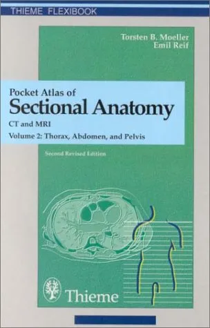
Pocket atlas of sectional anatomy: computed tomography and magnetic resonance imaging
4.5
Reviews from our users

You Can Ask your questions from this book's AI after Login
Each download or ask from book AI costs 2 points. To earn more free points, please visit the Points Guide Page and complete some valuable actions.Related Refrences:
Introduction to 'Pocket Atlas of Sectional Anatomy: Computed Tomography and Magnetic Resonance Imaging'
The 'Pocket Atlas of Sectional Anatomy: Computed Tomography and Magnetic Resonance Imaging' is an essential guide for medical professionals, radiologists, and students seeking comprehensive insights into the cross-sectional anatomy of the human body. This book, renowned for its authoritative content and illustrative clarity, serves as an invaluable reference for understanding anatomical structures as visualized through CT and MRI imaging.
Detailed Summary
The 'Pocket Atlas of Sectional Anatomy' offers a detailed exploration of the human anatomy using cutting-edge imaging technologies. Divided into meticulously organized sections, this atlas provides a structured overview of the head and neck, thorax, abdomen, pelvis, and extremities. Each section includes high-quality images that are complemented by precise labeling and detailed descriptions, making complex anatomical details more accessible and understandable.
The book emphasizes the correlation between anatomical structures and their appearance in CT and MRI images, offering clear, full-color illustrations along with concise insights into anatomical variations and pathologies. The authors, Torsten B. Moller and Emil Reif, have meticulously crafted this atlas to serve not only as a visual guide but also as an educational tool that deepens one's understanding of anatomy through cross-sectional imaging.
Key Takeaways
- Comprehensive Coverage: The atlas covers all major regions of the body, ensuring no aspect of sectional anatomy is overlooked.
- User-Friendly Format: Its pocket-sized format makes it a convenient resource for quick reference, both in clinical settings and educational environments.
- High-Quality Imaging: Featuring more than 900 high-resolution images, the book provides detailed visualization that enhances the learning experience.
- Clinical Relevance: Offers insights into clinical applications, facilitating an understanding of how anatomical knowledge is applied in diagnostics and treatment.
Famous Quotes from the Book
"Understanding sectional anatomy is pivotal to mastering the diagnostic arts of computed tomography and magnetic resonance imaging."
"The clarity of our anatomical understanding determines the clarity of our clinical decisions."
Why This Book Matters
In the ever-evolving landscape of medical imaging, the 'Pocket Atlas of Sectional Anatomy' stands out as a pivotal educational resource. It bridges the gap between theoretical knowledge and practical application, equipping readers with the ability to interpret complex imaging data with confidence. This book is of particular importance to radiologists, medical students, and healthcare professionals who rely on accurate anatomical knowledge for diagnosing and treating patients.
By offering a detailed examination of sectional anatomy through high-resolution CT and MRI images, this atlas enhances the reader's ability to correlate clinical findings with imaging results, fostering a deeper understanding that is crucial for effective patient care. Furthermore, its concise format and comprehensive coverage make it an ideal tool for both learning and review, ensuring that essential anatomical concepts are always within reach.
Free Direct Download
You Can Download this book after Login
Accessing books through legal platforms and public libraries not only supports the rights of authors and publishers but also contributes to the sustainability of reading culture. Before downloading, please take a moment to consider these options.
Find this book on other platforms:
WorldCat helps you find books in libraries worldwide.
See ratings, reviews, and discussions on Goodreads.
Find and buy rare or used books on AbeBooks.
![Essentials of Human Anatomy and Physiology [RENTAL EDITON]](https://s3.refhub.ir/images/thumb/Essentials_of_Human_Anatomy_and_Physiology__R_2981_uBSkHXG.webp)
