📚 چرا کتاب فیزیکی بخریم؟
- ✅ کیفیت چاپ عالی و کاغذ مرغوب
- ✅ راحتی مطالعه بدون نیاز به دستگاه
- ✅ امکان یادداشتبرداری و هایلایت
- ✅ تحویل سریع در سراسر کشور
قیمت نهایی کتاب
1,263,000 تومان
شامل چاپ و صحافیلذت ورق زدن یک کتاب واقعی
- سال انتشار: 2021
- صفحات: 1098
- اندازه فایل: 206 MB
- زبان: English
- انتشارات: ELSEVIER - HEALTH SCIENCE
- تعداد مشاهده: 374
- امتیاز کاربران: 4.5
-
شابک/ISBN:
9780323796651
0323796656
کلمات کلیدی:

Generate PowerPoint Files with LLM
⚡️SlideGenie is an intelligent educational slide generator (Open Source Repository) that leverages the power of OpenAI API to create engaging presentations and converts Mermaid diagrams into visual assets.
Features:
- AI-powered slide content generation
- Automatic Mermaid diagram to image conversion
- Customizable slide templates
- Multi-language support
- Educational content optimization
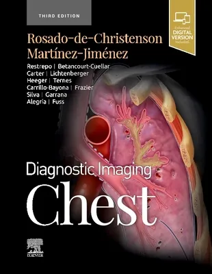
DIAGNOSTIC IMAGING: CHEST
4.5
بر اساس نظر کاربران

شما میتونید سوالاتتون در باره کتاب رو از هوش مصنوعیش بعد از ورود بپرسید
هر دانلود یا پرسش از هوش مصنوعی 2 امتیاز لازم دارد، برای بدست آوردن امتیاز رایگان، به صفحه ی راهنمای امتیازات سر بزنید و یک سری کار ارزشمند انجام بدین


معرفی کتاب
کتاب "DIAGNOSTIC IMAGING: CHEST" یکی از آثار برجسته در زمینه تصویربرداری تشخیصی سینه است که توسط MELISSA L MARTINEZ JIMENEZ SANTIAGO ROSADO-DE-CHRISTENSON نگاشته شده است. این کتاب به طور جامع و دقیق، تکنیکها، اصول و مبانی مهم در تصویربرداری پزشکی با تمرکز بر ناحیه سینه را پوشش میدهد.
خلاصهای جامع از کتاب
این اثر به طور کلی به بخشهای مختلفی تقسیم شده است که هر کدام به بررسی مفصل یک جنبه خاص از تصویربرداری سینه میپردازد. محتوای کتاب شامل توضیحات تکنیکی در مورد نحوه انجام صحیح CT Scan، MRI، و دیگر تکنیکهای موجود است. همچنین، کتاب به بررسی موارد مختلف بیماریها و شرایطی میپردازد که میتوانند در ناحیه سینه مشاهده شوند.
هدف این کتاب فراهم کردن منبعی معتبر و کارآمد برای پزشکان و متخصصین رادیولوژی است تا با استفاده از این اطلاعات، بتوانند به نتایج دقیقتری در تشخیص دست یابند و بیماران خود را بهتر درمان نمایند.
نکات کلیدی
- بررسی جامع از فنون مختلف تصویربرداری مانند CT و MRI.
- توضیحات واضح دربارهی تفسیر تصاویر به دست آمده از سینه.
- نمونههای متعدد از موارد بالینی واقعی برای درک بهتر مطالب.
- نکات مهم در زمینه کاهش خطرات رادیولوژیک در تصویربرداری پزشکی.
- راهکارهایی برای بهبود دقت در فرایند تصویربرداری و تشخیص.
جملات معروف از کتاب
“تصویربرداری سینه نه تنها نیازمند تخصص است، بلکه هنر تفسیر صحیح آن نیز اهمیت وافر دارد.”
“هر تصویر میتواند داستانی از وضعیت سلامتی بیمار را روایت کند، به شرط آنکه تفسیر صحیحی از آن به دست آید.”
اهمیت این کتاب
این کتاب به عنوان یک منبع جامع، برای پزشکان و متخصصین رشته رادیولوژی بسیار حیاتی است. با توجه به پیشرفتهای سریع در تکنولوژی تصویربرداری، درک صحیح و بهروز از این تصاویر میتواند نقش بزرگی در بهبود تشخیص و درمان بیماران ایفا کند.
کتاب "DIAGNOSTIC IMAGING: CHEST" با ارائه دادهها و تحلیلهای دقیق، به پزشکان این امکان را میدهد که خطاهای تشخیصی را کاهش داده و دقت عملکرد خود را افزایش دهند. در نهایت، این کتاب به عنوان مرجعی مهم میتواند به بهبود کیفیت خدمات بهداشتی و درمانی منجر شود.
Introduction
Welcome to the comprehensive resource for understanding thoracic imaging: "Diagnostic Imaging: Chest." This book endeavors to equip both novice and experienced radiologists with the knowledge required to navigate the complexities of thoracic imaging effectively.
Detailed Summary of the Book
In "Diagnostic Imaging: Chest," we delve into the intricate world of thoracic imaging with a comprehensive approach that covers a broad spectrum of chest pathologies. The book is structured to provide a systematic framework for the accurate diagnosis of various chest diseases using imaging technologies such as X-rays, computed tomography (CT), and magnetic resonance imaging (MRI).
The content begins with a detailed overview of normal chest anatomy and physiology as visualized on different imaging modalities. This foundation is crucial for understanding pathological deviations. Next, the book covers a wide range of conditions affecting the chest, including pulmonary, cardiovascular, and mediastinal diseases, along with infections, neoplasms, and interstitial lung diseases. Each section systematically addresses pathophysiology, imaging modalities, characteristic imaging features, and differential diagnosis.
One of the key features of this book is the extensive use of high-quality images that illustrate typical and atypical presentations of diseases, complemented by detailed legends that enhance understanding. Readers are guided through complex topics via organized headings, bullet points for clarity, and key points summaries that emphasize important concepts.
Key Takeaways
- Detailed coverage of thoracic imaging techniques and their applications.
- Expansive collection of high-resolution images for visual reference.
- Insightful discussions on the latest advancements in imaging technology.
- Comprehensive case studies that highlight real-world applications and challenges.
- Systematic approach to differential diagnosis and disease characterization.
Famous Quotes from the Book
“A picture can portray a multitude of findings that no amount of words can succinctly capture, especially in the realm of diagnostic imaging.”
“Successful radiological interpretation hinges on a meticulous understanding of anatomy and pathology, synthesizing them into a coherent narrative.”
Why This Book Matters
Diagnostic imaging has become pivotal in the management and treatment of chest diseases, rendering this book an invaluable resource for healthcare professionals. A precise and early diagnosis informed by imaging results can significantly influence patient outcomes, especially in complex conditions such as lung cancer or interstitial lung diseases.
This book not only aids radiologists but also serves as an essential guide for pulmonologists, thoracic surgeons, and other healthcare providers involved in managing chest diseases. Its detailed and organized layout allows for easy reference, promoting efficient learning and application in clinical practice. By offering updated information on recent advancements and techniques, "Diagnostic Imaging: Chest" enhances the practitioner’s capability to deliver evidence-based care.
In a field as dynamic and critical as thoracic radiology, staying abreast of evolving methodologies and having access to a robust reference guide is crucial. This book fulfills that need, making it a critical addition to the libraries of medical professionals committed to improving patient care.
دانلود رایگان مستقیم
شما میتونید سوالاتتون در باره کتاب رو از هوش مصنوعیش بعد از ورود بپرسید
دسترسی به کتابها از طریق پلتفرمهای قانونی و کتابخانههای عمومی نه تنها از حقوق نویسندگان و ناشران حمایت میکند، بلکه به پایداری فرهنگ کتابخوانی نیز کمک میرساند. پیش از دانلود، لحظهای به بررسی این گزینهها فکر کنید.
این کتاب رو در پلتفرم های دیگه ببینید
WorldCat به شما کمک میکنه تا کتاب ها رو در کتابخانه های سراسر دنیا پیدا کنید
امتیازها، نظرات تخصصی و صحبت ها درباره کتاب را در Goodreads ببینید
کتابهای کمیاب یا دست دوم را در AbeBooks پیدا کنید و بخرید

1374
بازدید4.5
امتیاز0
نظر98%
رضایتنظرات:
4.5
بر اساس 0 نظر کاربران
Questions & Answers
Ask questions about this book or help others by answering
No questions yet. Be the first to ask!