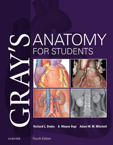
Gray 39's Anatomy for Students
Richard L. Drake,A. Wayne Vogl,Adam W. M. Mitchell (eds.)
2020
View book
Book guide and evaluation
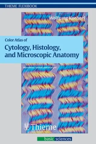
Wolfgang Kuehnel
0 reviews
Published
pages
views
Introduction Welcome to the "Color Atlas of Cytology, Histology, and Microscopic Anatomy," a comprehensive resource meticulously designed to cater to the needs of students, researchers, and professionals in the fields of biology, medicine, and related sciences. This book s
Before you read
Welcome to the "Color Atlas of Cytology, Histology, and Microscopic Anatomy," a comprehensive resource meticulously designed to cater to the needs of students, researchers, and professionals in the fields of biology, medicine, and related sciences. This book serves as both an instructional guide and a reference point, bridging the gap between academic learning and practical application in cytology and histology.
The "Color Atlas of Cytology, Histology, and Microscopic Anatomy" is structured to provide a clear, coherent, and detailed exploration into the microscopic world. The atlas offers an extensive collection of richly colored, high-quality images that depict a wide array of cell types, tissues, and microscopic structures. These images are paired with concise descriptions that elucidate the function, location, and importance of each anatomical feature. Throughout the book, readers will find a systematic breakdown beginning with cells—the fundamental units of life—progressing to tissues, and culminating in comprehensive discussions of complex anatomic structures.
The book is divided into well-defined sections that start with cellular biology and advance through epithelial tissues, connective tissues, muscular and nervous systems, and then delve into organ systems. Each section is crafted to offer insights into the appearance, structure, and relationships between different microscopic anatomic entities. This organization not only improves the reader’s understanding but also aids in retaining critical information, making it an invaluable educational tool.
"The microscopic world is as vast and intricate as the universe itself. To understand it is to appreciate the profound complexity of life."
"Each cell, no matter how small, carries the story of life, a mosaic of biology waiting to be explored under the lens."
The "Color Atlas of Cytology, Histology, and Microscopic Anatomy" is more than just a collection of images and descriptions; it is a tool that empowers learning and fosters deeper appreciation for microscopic anatomy. In medical and scientific education, understanding the microanatomy of cells and tissues is crucial for diagnosing diseases, developing treatments, and advancing biological sciences. This atlas stands as a vital aid in cultivating the analytical skills needed to explore these microscopic structures effectively.
Moreover, the book is designed to cater to a diverse audience, fulfilling the requirements of students embarking on their educational journey in health sciences, as well as professionals seeking a reliable reference in their field. By leveraging the power of visually driven learning paired with precise explanations, this atlas bridges the gap between theory and application, highlighting the relevance of microanatomy in real-world contexts.
Ultimately, whether for educational purposes or professional use, this book is essential for anyone looking to delve into the fascinating intricacies of the microscopic world and gain a holistic understanding of life's foundational structures.
Your question is answered in the context of this title and author. Each answer uses 2 points.
0 reviews, 4.7 average out of 5
Sign in to publish a review.
Ask a focused question and learn from the community.
Related references that continue this learning path.
Richard L. Drake,A. Wayne Vogl,Adam W. M. Mitchell (eds.)
Klaus Wolff,Lowell Goldsmith,Stephen Katz,Barbara Gilchrest,Amy Paller,David Leffell